Fertilization – the beginning of a new life
New life begins with the meeting of the female and male reproductive cells – the ovum and the sperm. Both the female and male germ cells must undergo a long process of development to reach full maturity.
Sperm formation takes about 70 days. They are produced in the testes, which are located outside the body because excessively high temperatures negatively affect spermatogenesis. The vas deferens and epididymis store the sperm until ejaculation. The prostate gland and seminal vesicles secrete fluids that nourish and protect the sperm; this fluid, together with the sperm cells, is called semen. The secretions from each of these glands are alkaline, creating a suitable environment for the sperm to survive and protecting them from potential damage.
The growth and maturation of the ovarian follicle containing the egg is a cyclical process that is repeated in each menstrual cycle. Each female child, already during fetal development, is endowed with a certain number of primary follicles deposited in the ovaries (the so-called ovarian reserve). However, in each cycle, typically only one of these follicles reaches the stage of a Graafian follicle.
Ovaries measure approximately 2.5 to 5 cm in length and 1.5 to 3 cm in width. Their function is essential for human fertility, namely the release of a mature germ cell. The primary follicles are located closest to the ovarian surface. Above them are the growing follicles, which are more sensitive to follicle-stimulating hormone (FSH) and secrete estradiol and androgens. Through the process of natural selection among the growing follicles, one follicle (in exceptional cases more than one) is chosen to mature into a Graafian follicle and release the egg during ovulation.
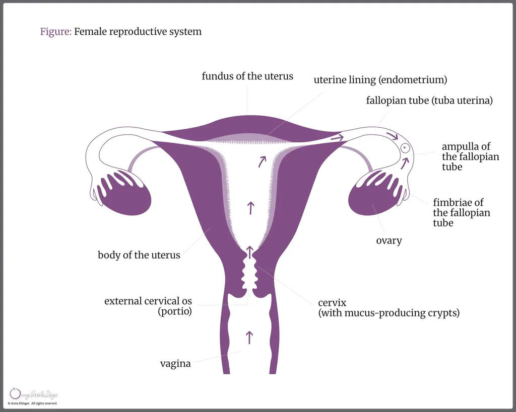
The sperm begins its journey toward the egg inside the vagina. The vagina is a musculomembranous duct that is highly extensible and elastic. It measures 6 to 12 cm in length and has an acidic environment that is generally hostile to sperm. During the fertile period, the vaginal pH changes due to the secretion of alkaline cervical mucus, which helps the sperm survive. After intercourse, the sperm collect in the vaginal vault and then ascend through the cervix. Immediately upon entering the vagina, sperm are incapable of fertilizing an egg. To acquire this capacity, they must undergo the capacitation process in the female reproductive tract, which takes approximately seven hours.
The cervix is a narrow canal, 2–2.5 cm long, with its external opening leading to the vagina and its internal opening connecting to the uterine cavity. Within the cervical crypts, columnar cells produce different types of mucus. An increasing concentration of estradiol triggers the production of estrogenic mucus, which facilitates the entry of sperm into the reproductive system, whereas an increase in progesterone secretion thickens the cervical mucus and prevents sperm from entering the uterus.
During the periovulatory phase, the cervical canal opens, facilitating sperm penetration. In addition, the glucose present in cervical mucus provides an ideal nutrient for sperm, supporting their final stage of maturation. The presence of mucus also helps select healthy sperm and block defective or abnormal ones.
Ovulation is the culminating event of each menstrual cycle. It is preceded (by approximately 36 to 42 hours) by a sharp surge in luteinizing hormone (LH), which is necessary for ovulation. The peak of LH secretion occurs about 17 hours before ovulation, while the peak of estrogen secretion occurs about 24 hours before ovulation. The mature follicle, ready to rupture, measures approximately 2.0 to 2.5 cm in diameter. The egg released from the Graafian follicle is captured by the fimbriae of the fallopian tube and guided into its internal portion.
The fallopian tubes are 14-20 cm long and connect the ovaries to the uterus. The first section of the tube is the infundibulum, followed by the ampullary region – the widest part, where sperm meet the egg. In the fallopian tube, sperm can remain viable for several days while awaiting the egg’s final maturation. The acrosome reaction occurs near the oocyte, triggered by substances released from the egg. One sperm penetrates the egg, its head swells, and it becomes a typical cell nucleus. This process results in the formation of a fertilized egg – the zygote. The egg’s metabolism is likely activated by factors supplied by the sperm, which also contribute to the hereditary features of the father, complementing those of the mother. The embryo is programmed to travel through the tube over a defined period (5–6 days) and to implant in the uterus.
The uterus is a muscular organ, 7-9 cm in length. Its lining, called the endometrium, responds strongly to sex hormones and undergoes cyclical changes during the menstrual cycle. After ovulation, the endometrium transforms to facilitate embryo implantation, which typically occurs about seven days after fertilization.
During fetal development, the primary function of the uterus is to protect the fetus and mediate the delivery of nutrients.
The remains of the ruptured Graafian follicle form the corpus luteum, which, under the influence of luteinizing hormone (LH), secretes progesterone. The developing human embryo (trophoblast) begins to produce its own hormone – human chorionic gonadotropin (hCG) – which stimulates the corpus luteum to secrete even more progesterone. The corpus luteum continues its function as the corpus luteum of pregnancy, nourishing the embryo and then the fetus, until the placenta is fully developed, typically around 16 weeks.
When the fetus is ready for birth, the uterus begins rhythmic contractions to expel the child, followed by the separation of the placenta from the uterine wall. After delivery, the uterus shrinks and undergoes involution during the postpartum period, which lasts 5 to 8 weeks.
Thus, after approximately 266 days of pregnancy (according to Döring’s research), a new person is born.